
Nuclear Scintigraphy, or bone scanning, pinpoints areas of bone and in some cases soft tissue pathology due to injury. It is used, most commonly, in difficult lameness investigations to detect specific regions of the limbs that are resulting in lameness.
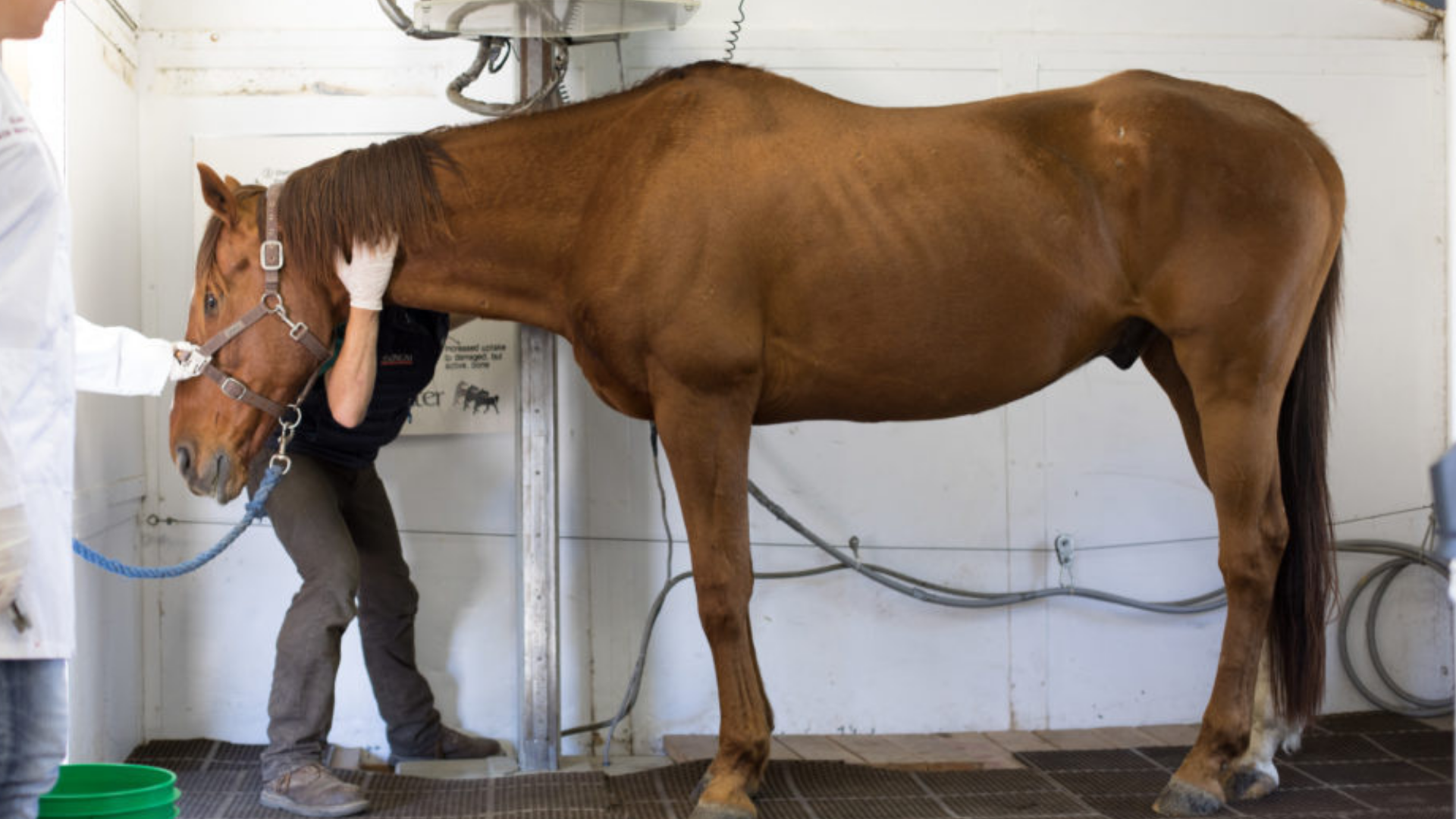
The procedure is performed standing, with the horse sedated to keep him/her as still as possible. A radioactive drug is injected into the jugular vein through a pre-placed venous catheter. The radioactive drug is combined with a special marker that binds to bone. The bone marker and radioactive isotope combination then distributes through the body via the blood stream and eventually reaches the bone.
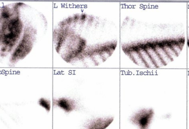
The drug then binds to the bone. Bone that is damaged has an increased blood supply and thus more of the drug will be taken up in such areas. A gamma camera is then used to detect the radioactivity, sequentially, at specific locations on the body and limbs and then software converts the data to a picture on a computer. The resultant images show areas of increased radiopharmaceutical uptake or “hot spots” on the body that highlight where the injury is located.
When should I opt for a bone scan for my horse?
A bone scan is a good option when you are dealing with a difficult or multi-focal lameness. The bone scan can tell you all locations that are affected – both primary and secondary issues.
- Difficult to block lamenesses
- Hind end issues
- Transient lamenesses
- Horses lame in multiple limbs
- Chronic issues
- Prepurchase exam
- Can diagnose infections, tumors and arthritis
- Earlier detection means earlier treatment and higher success rates
- Find performance limiting issues
- Good for subclinical complaints
Nuclear scintigraphy is safe for your horse and is a relatively short procedure lasting only a few hours. The radioactive isotope is benign. The isotope decays 97% in 30 hours, so horses are able to leave the day following the procedure.
Nuclear Scintigraphy is a non invasive technique (it does not require surgery). It complements but does not replace survey radiography. The changes noted in scintigraphy usually precede the changes noted on x-rays, since the bone’s metabolism usually changes before the bone’s structure changes. Scintigraphy is useful in the evaluation of patients with poorly localized lameness, especially when x-rays are inconclusive. Small lesions can result in significant changes in the metabolism of the bone which are assessed through the images. Bone scans are useful for diagnosing conditions such as infections, tumors and arthritis. Earlier diagnosis means earlier treatment and increases the probability that the treatment will be more successful. Nuclear Scintigraphy offers specific diagnosis of injuries that are seriously performance limiting often before they become seriously damaging.