
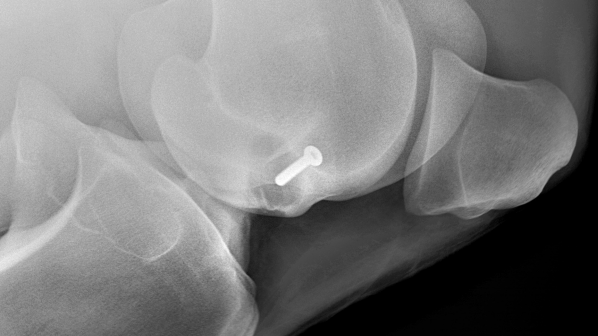
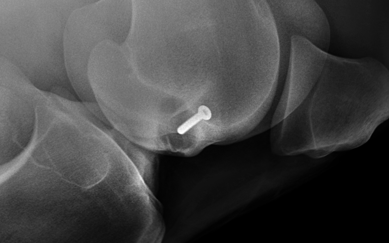
Radiographs are often the first diagnostics imaging modality used to evaluate lameness. Digital Radiography allows for potentially greater detail than conventional radiography by permitting each digitized image to be processed by the computer.
The state-of-the-art digital radiography systems at The Equine Center are powerful, high speed x-ray machines that make it possible to image difficult body parts such as the neck, chest, and pelvis of large adult horses. The systems are portable and our veterinarians can take detailed, digital x-rays in the field and view the images within seconds. This greatly speeds up the diagnosis and treatment of your horse.
WHAT CAN DIGITAL RADIOGRAPHY BE USED TO DIAGNOSE?
The Equine Center provides digital radiography services for equine patients both in our hospital and on the farm.
- Navicular disease
- Osteoarthritis/degenerative joint disease
- Fractures
- Podiatry aid
- Laminitis
- Osteochondritid dissecans
- Wounds – foreign bodies, penetration of joints
- Prepurchase exam survey
- Venogram for blood flow analysis in feet
Radiographs are the work-horse of diagnostic imaging. Today’s portable machines are easy to use and produce high-resolution images that can be reviewed instantly on a laptop. It’s simple to send images electronically for evaluation and in some cases, a second opinion from board-certified radiologists.
In collaboration with our Farrier team we provide corrective trimming and shoeing through radiographic guidance. Radiographs are taken for a hoof evaluation; specific software determines the angles and balance of the hoof and phalanges. After assessment, we work with the farrier to correct the condition by trimming and/or shoeing the horse.