
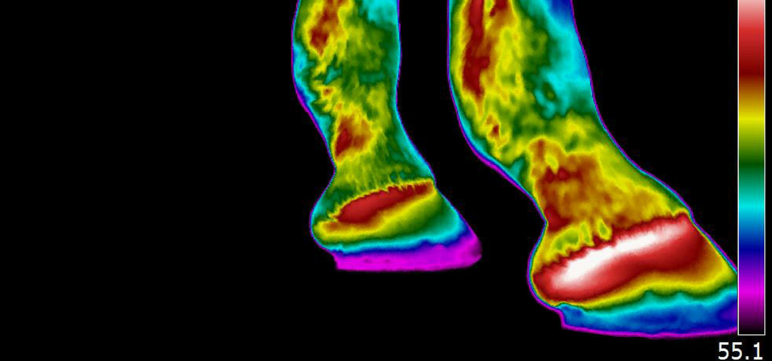
Thermography is a great non-invasive physiological imaging tool that can detect areas of inflammation and changes in blood flow in the horse. Equine infrared imaging can detect musculo-skeletal injuries several weeks before they are visually detectable.
So how does thermography work? The thermography camera detects infrared waves on the body surface that are invisible to the human eye and converts them to an image we can see. Consider this: You bang your knee; the area becomes hot, red, and inflamed; and at a cellular level as an immune response, the body releases chemicals such as histamine. Changes in blood flow might directly correlate with inflammation. Thus, at the most basic level, where there is increased circulation there might be inflammation (becoming warmer). The opposite is also true: With chronic disease, scarring, atrophy (muscle wasting/loss), nerve damage, or disuse, areas might become cooler.
WHAT CONDITIONS ARE DETECTABLE WITH THERMOGRAPHY?
Performance limiting or career-ending conditions can be detected including:
- Tendon/ligament problems
- Kissing spine
- Soreness from poorly fitting saddles
- Saddle fit evaluation
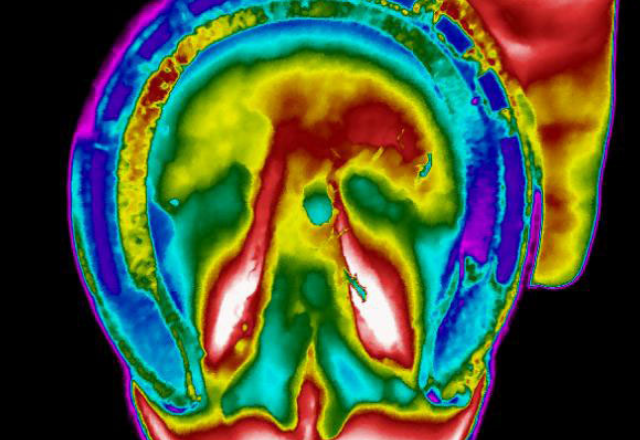
- Hoof abnormalities
- Arthritis
- Cervical issues
- Cold dermatomes/Pinched nerves
- Prepurchase evaluation
- Sub-clinical inflammation affecting performance
The thermal images may indicate the onset of inflammatory reaction in joints, ligaments and tendons from two to six weeks prior to clinical symptoms (lameness, swelling etc).
Symmetry is key when interpreting images. By comparing a horse’s parts left-to-right, the individual patient serves as its own control. Also consider that a horse with an injured right front flexor tendon, for example, might be offloading from the sore leg to the opposite (left front) limb or even the hind end. With a whole horse scan, veterinarians can detect these compensatory problems, begin treatment, and prevent further injury.